Clinical diagnosis
A clinical examination will assess the general health of your dog and establish whether the problem is neurological. There are many possible causes of back pain and a disc extrusion cannot be diagnosed on the basis of a clinical examination alone.
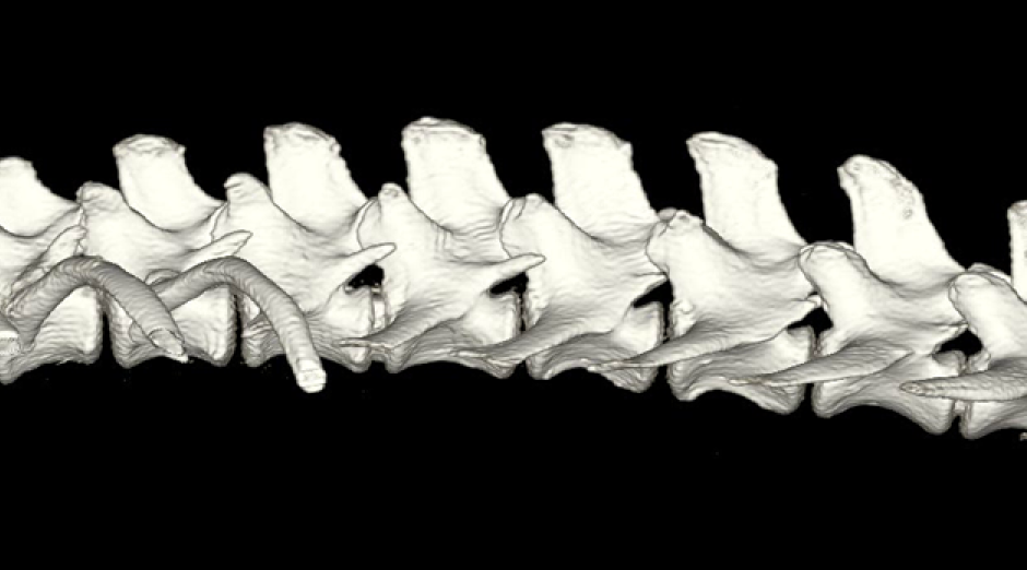
Spinal cord compression caused by disc extrusion cannot be diagnosed by X-rays. This can be done by Magnetic Resonance Imaging (MRI), Computed Tomography (CT) or Myelography.
- An MRI scan provides cross-sectional images of the spinal cord and discs
- A CT scan is, effectively, a 3D X-ray which, with computer processing also allows the soft tissues to be seen
- Myelography uses a dye injected into the spinal column that shows up on X-rays and can show cord compression
In general, MRI is the preferred diagnostic imaging technique as the results are much clearer and easier to interpret. It is, however, the most expensive diagnostic option. MRI and CT are both safe diagnostic techniques but your dog will probably need to be referred to a specialist for these. There are some risks associated with Myelography because it requires an injection into the spine and it is less likely to be recommended now, although it may still be a viable lower-cost option.
It is unlikely that your dog will need to be referred to have an MRI or CT scan if it has been graded as 1 or 2, where conservative treatment is the usual starting point.
It is important to get the right diagnosis, quickly. There is information on other possible (non-IVDD) diagnoses, here.
Depending on your dog's symptoms and the results of any imaging, your vet may recommend conservative or surgical treatments. The likely success of a course of treatment depends on the severity of symptoms and how quickly treatment can begin.
Here's our checklist of 10 questions to ask your vet.
Diagnostic imaging and surgical treatment will be expensive, maybe £6,000 to £10,000 if you need both an MRI and surgery carried out by a referral practice. We strongly recommend you ensure you have adequate insurance cover or other ways to pay for your dog's treatment. Make sure you discuss the available range of options and costs with your vet before being rushed into a decision on diagnosis and treatment.
Open Access Paper: Diagnostic Imaging in Intervertebral Disc Disease (Front. Vet. Sci., 22 October 2020)
Dr Marianne Dorn has provided a downloadable guide on how best to help the dachshund presenting with back disease. It’s written for first-opinion vets in practice and can be found in the March 2019 edition of In Practice journal.
A pdf version of this paper may be downloaded for educational purposes by clicking here
To cite the attached article: “Dorn, M., 2019. First-opinion approach to the dachshund with intervertebral disc herniation. In Practice, 41(2), pp.59-68″
Which discs are most at risk?


